Motion Compensation of Optical Coherence Tomographs

TNT members involved in this project:
Prof. Dr.-Ing. Bodo Rosenhahn
Dr.-Ing. Dipl. Math. Oliver Müller
Show active Staff only

Optical Coherence Tomography1 (OCT) is a noninvasive high resolution imaging technique which is used here for in vivo 3D volume capturing of mouse skin tissue (hairless mouse) around a percutaneous implant. 
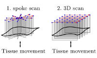
3D volume acquisition in OCT is enabled by using a lateral scanning scheme. The scanning process can take several seconds. During this time period, the skin tissue has to stay fixed in order to avoid motion artifacts. A custom made animal positioning table was used in order to fixate the mouse during in vivo OCT acquisition. 
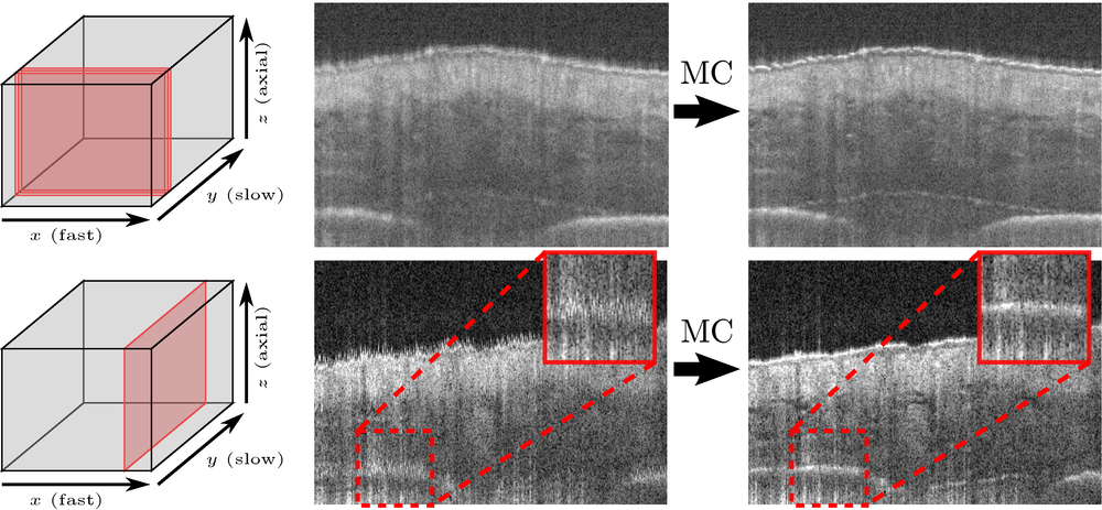
Nevertheless, slight up and down movements introducing motion artifacts in the OCT data, caused by heart beating and breathing of the mouse, could not be avoided (see Fig. 1).

Percutaneous Implant
Figure 1: In vivo OCT scan and its motion compensated result. Top: Fast scanning direction (three consecutive B-scans averaged) Bottom: Reslice along the slow scanning direction.
The goal of the project is to develop a fully automatic motion compensation algorithm. The method should work without using a reference measure. In order to reliably estimate the tissue motion during OCT acquisition, several OCT scans of the same tissue region using different scanning schemes are combined.

We present a probabilistic motion compensation algorithm using Conditional Random Fields (CRF). Simple motion compensation approaches using crosscorrelation of neighboring image columns tend to oversmoothing, i.e. flatten the tissue surface. The major problem here is in how to separate the motion shift (which lives in time domain) from the skin tissue structure change (spatial domain).
Random Field Decomposition

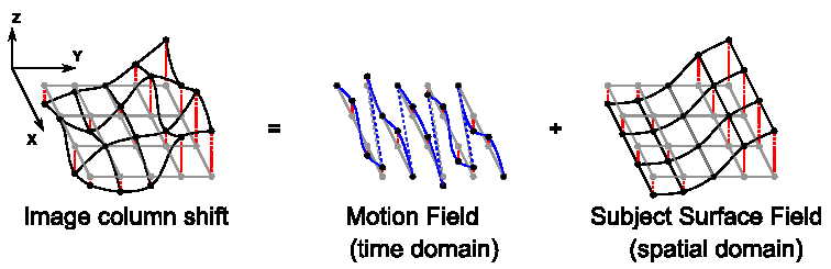
In our approach, we deal with this problem by introducing two CRF energy terms:

  1. Intra-scanning scheme energy term - treat each scanning scheme separately by using crosscorrelations of neighboring image columns within a scanning scheme.
  2. Inter-scanning scheme energy term - combine different scanning schemes by using crosscorrelations of image columns at the same spatial position across different scanning schemes.
Additionally, a regularizing term is added in order to retrieve a smooth motion field along the time axis.

Our experiments are done using two scanning schemes: a spoke pattern and a dense 3D scanning scheme. Even though using this limited set of scanning schemes, we are able to significantly reduce motion artifacts while preserving the tissue surface geometry.


  • Conference Contributions
    • Oliver Müller, Sabine Donner, Tobias Klinder, Ivonne Bartsch, Alexander Krüger, Alexander Heisterkamp, Bodo Rosenhahn
      Compensating motion artifacts of 3D in vivo SD-OCT scans
      Medical Image Computing and Computer Assisted Intervention (MICCAI), Vol. 7510, pp. 198--205, October 2012, edited by Nicholas Ayache, Hervé Delingette, Polina Golland, Kensaku Mori
  • Journals
    • Sabine Donner, Oliver Müller, Frank Witte, Ivonne Bartsch, Elmar Willbold, Tammo Ripken, Alexander Heisterkamp, Bodo Rosenhahn, Alexander Krüger
      In situ optical coherence tomography of percutaneous implant-tissue interfaces in a murine model
      Biomedical Engineering/Biomedizinische Technik, De Gruyter, pp. 1-9, Karlsruhe, May 2013, edited by Dössel, Olaf