Undistortion of Optical Coherence Tomographs of Percutaneous Implants

TNT members involved in this project:
Prof. Dr.-Ing. Bodo Rosenhahn
Dr.-Ing. Dipl. Math. Oliver Müller
Show active Staff only

Optical Coherence Tomography1 (OCT) is a newly established noninvasive imaging technique which is used here for in vivo biocompatibility studies of percutaneous2 implants. A drawback of this technique is that the OCT scans are optical distorted. The distortion mostly depends on the optical refraction within the x-rayed material. Refraction correction of the OCT scans is an essential step towards further tasks like morphometric analysis of the implant--tissue interface. As modern OCT scanners are capable for near realtime image capturing with high image resolutions and therefore generate huge amounts of data, methods for automatic image processing are required.

Percutaneous Implant OCT Undistortion
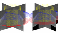
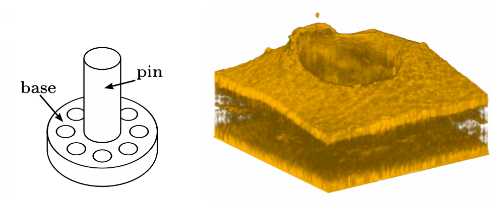
Figure 1: Model of a percutaneous implant and volume rendering of a skin tissue (the hole indicates the position of the implant pin) Figure 2: 3D segmentation (blue: skin surface, red: base contour, yellow: pin) and refractive undistortion

The goal of this project is to provide a fully automatic refractive undistortion framework of the OCT scans using model based 3D segmentation.

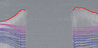
The first step involves the segmentation of the skin and percutaneous implant base using Markov random fields and a generalized Hough transform approach. 
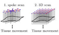
In the second step, the amount of image distortion is estimated using a refractive distortion model. Model parameters are estimated by incorporating a-priori knowledge of the planar shape of the percutaneous implant base. Finally, the images are undistorted according to the refractive distortion model and the previously estimated model parameters.


In Dec. 4, 2011 our MICCAI article appeared as feature of the week in OCT-news!

  • Conference Contributions
    • Oliver Müller, Sabine Donner, Tobias Klinder, Ivonne Bartsch, Alexander Krüger, Alexander Heisterkamp, Bodo Rosenhahn
      Compensating motion artifacts of 3D in vivo SD-OCT scans
      Medical Image Computing and Computer Assisted Intervention (MICCAI), Vol. 7510, pp. 198--205, October 2012, edited by Nicholas Ayache, Hervé Delingette, Polina Golland, Kensaku Mori
    • Oliver Müller, Sabine Donner, Tobias Klinder, Ralf Dragon, Ivonne Bartsch, Frank Witte, Alexander Krüger, Alexander Heisterkamp, Bodo Rosenhahn
      Model Based 3D Segmentation and OCT Image Undistortion of Percutaneous Implants
      Medical Image Computing and Computer-Assisted Intervention – MICCAI 2011 14th International Conference, Lecture Notes in Computer Science (LNCS), Springer Berlin / Heidelberg, Vol. 6893, pp. 454-462, September 2011, edited by Fichtinger, Gabor and Martel, Anne and Peters, Terry
  • Journals
    • Sabine Donner, Oliver Müller, Frank Witte, Ivonne Bartsch, Elmar Willbold, Tammo Ripken, Alexander Heisterkamp, Bodo Rosenhahn, Alexander Krüger
      In situ optical coherence tomography of percutaneous implant-tissue interfaces in a murine model
      Biomedical Engineering/Biomedizinische Technik, De Gruyter, pp. 1-9, Karlsruhe, May 2013, edited by Dössel, Olaf